body structure
- (Body Structures) Parts of the body such as arms, legs; organs such as the heart or kidney and those systems which supports abilities.
- structure: a particular complex anatomical part of a living thing; “he has good bone structure”
golgi
- Golgi is a tiny lunar impact crater located in the Oceanus Procellarum, over 150 kilometers to the north of the crater Schiaparelli. It is a circular, cup-shaped impact formation with an interior albedo that is higher than the surrounding dark lunar mare.
- Camillo (1844–1926), Italian histologist and anatomist. He devised a staining technique to investigate nerve tissue, classified types of nerve cells, and described the structure in the cytoplasm of most cells, now named after him. Nobel Prize for Physiology or Medicine (1906, shared with Ramón y Cajal)
- Italian histologist noted for work on the structure of the nervous system and for his discovery of Golgi bodies (1844-1926)
- (in Camillo Golgi (Italian physician and cytologist))

golgi body structure – The Human
The Human Brain: Prenatal Development and Structure
This book is unique among the current literature in that it systematically documents the prenatal structural development of the human brain. It is based on lifelong study using essentially a single staining procedure, the classic rapid Golgi procedure, which ensures an unusual and desirable uniformity in the observations. The book is amply illustrated with 81 large, high-quality color photomicrographs never previously reproduced. These photomicrographs, obtained at 6, 7, 11, 15, 18, 20, 25, 30, 35, and 40 weeks of gestation, offer a fascinating insight into the sequential prenatal development of neurons, blood vessels, and glia in the human brain.
Dunaliella
Dunaliella: Cells mostly radially symmetrical, sometimes bilaterally symmetrical, flattened, dorsoventrally curved or slightly asymmetrical; cell shape ellipsoidal, ovoid, cylindrical, pyriform, or fusiform to almost spherical; cell size and shape may vary within a given species depending on different environmental conditions; cell surface smooth with distinctive mucilaginous cell coat; flagella 2, of equal length; chloroplast single, cup-, dish-, or bell-shaped; pyrenoid basal with continuous starch shell; eyespot anterior; nucleus anterior; Golgi bodies (2-4) parabasal; contractile vacuoles absent (freshwater species of doubtful affiliation); asexual reproduction by longitudinal division of vegetative flagellate cells; asexual cysts subspherical, thick-walled with bumpy surface; sexual reproduction isogamous, gametic fusion involves flagellar agglutination and activation of mating structures; several species are homothallic, the type species is reported to be heterothallic; zygote with a thick, smooth wall; after a resting stage, the zygote forms 32 progeny cells, which are liberated through a rupture in the zygote cell wall; meiosis takes place during germination of the zygote; habitat euryhaline or hypersaline. This is an un-named species of Dunaliella, a green alga and member of the phytoplankton that is often associated with hypersaline conditions. The chloroplast is bowl-shaped and fills the base of the cell. There is a yellowy eyespot to the left side of the plastid in this image. The nucleus with nucleolus lies near the front of the cell. There are two equally long flagella which beat in a breast-stroke motion. Phase contrast micrograph. D. J. Patterson, image used under license to MBL (micro*scope)
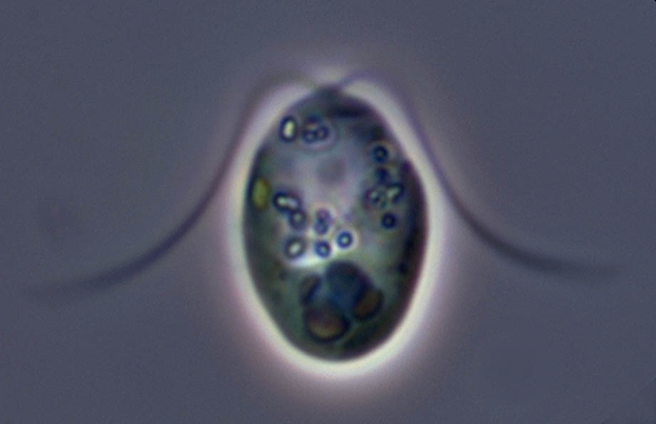
Dunaliella 2

Dunaliella: Cells mostly radially symmetrical, sometimes bilaterally symmetrical, flattened, dorsoventrally curved or slightly asymmetrical; cell shape ellipsoidal, ovoid, cylindrical, pyriform, or fusiform to almost spherical; cell size and shape may vary within a given species depending on different environmental conditions; cell surface smooth with distinctive mucilaginous cell coat; flagella 2, of equal length; chloroplast single, cup-, dish-, or bell-shaped; pyrenoid basal with continuous starch shell; eyespot anterior; nucleus anterior; Golgi bodies (2-4) parabasal; contractile vacuoles absent (freshwater species of doubtful affiliation); asexual reproduction by longitudinal division of vegetative flagellate cells; asexual cysts subspherical, thick-walled with bumpy surface; sexual reproduction isogamous, gametic fusion involves flagellar agglutination and activation of mating structures; several species are homothallic, the type species is reported to be heterothallic; zygote with a thick, smooth wall; after a resting stage, the zygote forms 32 progeny cells, which are liberated through a rupture in the zygote cell wall; meiosis takes place during germination of the zygote; habitat euryhaline or hypersaline.Dunaliella is a green alga and is a common member of the phytoplankton in salty water bodies. These cells were abundant in a collection taken at the margins of Mono lake (at Navy Beach). Each cell has a cup-shaped or bowl-shpaed chloroplast at the posterior end of the cell, aneriorly they have two equally long flagella. Differential interference contrast optics. D. J. Patterson, image used under license to MBL (micro*scope).
golgi body structure

The life of Camillo Golgi was an extraordinary intellectual adventure in three major fields of biology and medicine, namely neuroscience, emerging cell biology, and the new science of medical microbiology.
in 1873, Golgi published the description of a revolutionary histological technique which allowed one, for the first time, to visualize a single nerve cell with all its ramifications, and which could be followed and analyzed even at a great distance from the cell bodies. The so-called “black reaction” (later named the “Golgi method”) provided the spark to a truly scientific revolution which allowed the morphology and the basic architecture of the cerebral tissue to be evidenced in all its complexity, thus contributing to the foundation of modern neuroscience. It has been written that, in the same way Galileo Galilei was able to find new stars observing any sky region with his telescope. Golgi was able to find new nervous structures and nerve cells by applying his black reaction to any brain region. Finally, the details of the most complex structure in the known universe, the brain, could be characterized.
Golgi’s black reaction is just one of his many successes and contributions to society. As this book illustrates, he has also strongly contributed to the development of cell biology with the “internal reticular apparatus” (later called the “Golgi apparatus”) and to medical microbiology with his studies on malaria. Engrossing and authoritative, Golgi: A Biography of the Founder of Modern Neuroscience, is a detailed account of one of the most prominent European researchers of his time.
